
Anatomy of the Vestibular System
- What are the main components of the vestibular system?
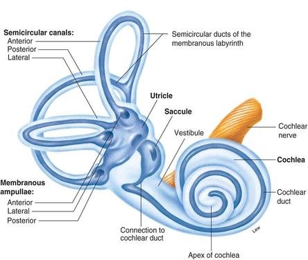
A: The vestibular system includes the semicircular canals, utricle, saccule, and the vestibular portion of the eighth cranial nerve (CN VIII). These structures detect head motion and position to maintain balance and spatial orientation.
2. What is the function of the semicircular canals?
A: The semicircular canals detect angular (rotational) movements of the head. Each canal corresponds to a different plane (horizontal, anterior, posterior), helping detect movement in all directions.
3. How many semicircular canals are there, and how are they oriented?
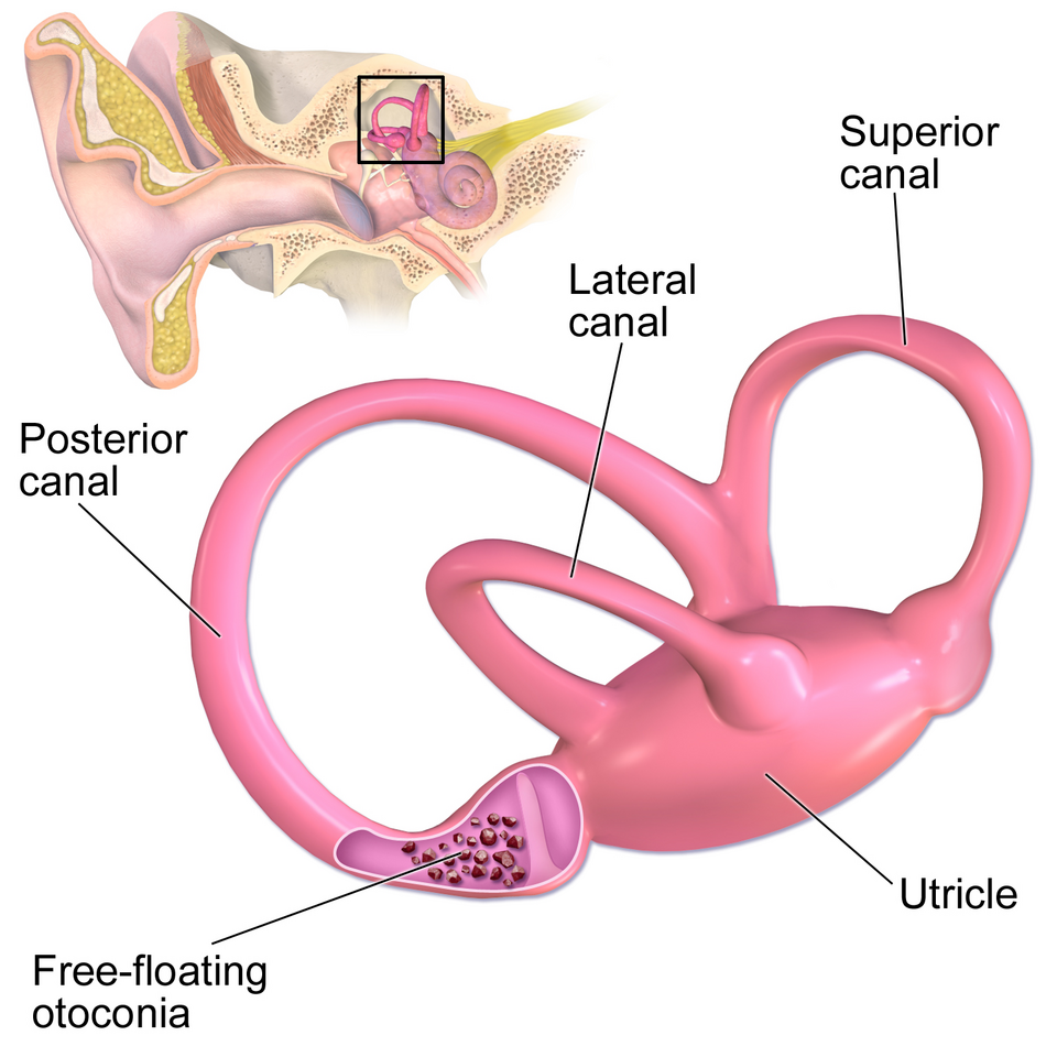
A: There are three semicircular canals per ear—horizontal (lateral), anterior (superior), and posterior. They are oriented roughly at right angles to each other, allowing detection of movement in three-dimensional space.
4. What is the ampulla, and what is its role?
A: Each semicircular canal (anterior, posterior, and horizontal) has one ampulla. Inside this structure, you will find two critical components:
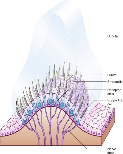
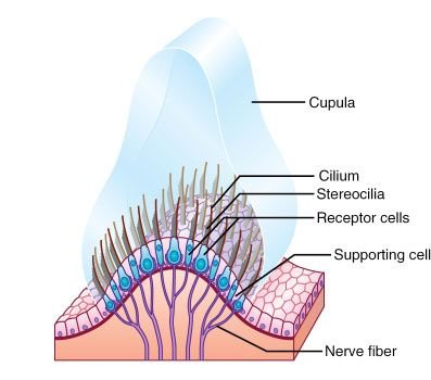
- Crista Ampullaris: A mound of sensory hair cells and supporting cells. Each hair cell features cilia (stereocilia and a single kinocilium) that project upward.
- Cupula: A gelatinous, sail-like structure that sits atop the crista ampullaris, completely spanning the width of the ampulla and embedding the cilia of the hair cells.
The Role of the Ampulla
The primary role of the ampulla is to convert the mechanical energy of fluid movement into neural signals for the brain, a process known as mechanotransduction.
Detection of Angular Acceleration
The ampulla is specifically designed to detect rotational movement (angular acceleration). When the head turns, the bone of the semicircular canal moves with it, but the endolymph (fluid) within the canal lags behind due to inertia.
2. Deflection of the Cupula
As the fluid lags, it pushes against the cupula. Because the cupula is flexible, it bends in the opposite direction of the head’s rotation.
3. Neural Signaling
- Excitation: If the cupula bends toward the kinocilium (the tallest hair), the sensory hair cells depolarize. This increases the firing rate of the vestibular nerve, signaling to the brain that the head is rotating in a specific direction.
- Inhibition: If the cupula bends away from the kinocilium, the cells hyperpolarize, decreasing the firing rate.
4. Maintaining Equilibrium
The brain integrates these signals from all six ampullae (three on each side of the head) to coordinate the Vestibulo-Ocular Reflex (VOR)—allowing your eyes to stay fixed on a target while your head moves—and to maintain postural balance.
5. What are the otolith organs, and what do they detect?
A: The otolith organs are two sensory structures in the inner ear—the utricle and the saccule—that form part of the vestibular system. While the semicircular canals detect rotation, the otolith organs are responsible for sensing gravity and movement in a straight line.
The Utricle and Saccule
Both organs contain a specialized sensory patch called the macula, which consists of hair cells embedded in a gelatinous layer.
- The Utricle: Positioned horizontally, it primarily detects horizontal linear acceleration (such as a car moving forward or side-to-side) and head tilts in the horizontal plane.
- The Saccule: Positioned vertically, it primarily detects vertical linear acceleration (such as the up-and-down motion of an elevator) and gravity.
Mechanism: How They Work
The term “otolith” literally means “ear stone,” which refers to the unique way these organs function:
1. The Otolithic Membrane
On top of the gelatinous layer in the macula sit thousands of small calcium carbonate crystals called otoconia (or otoliths). These crystals make the membrane significantly heavier than the surrounding fluid and structures.
2. Detection of Linear Acceleration
When you move forward or backward, the heavy otoconia lag behind due to inertia. This lag pulls the gelatinous layer, which in turn bends the cilia (hairs) of the sensory hair cells.
3. Detection of Static Tilt (Gravity)
When you tilt your head, gravity pulls on the heavy otoconia, causing them to shift. This provides the brain with constant information about the head’s position relative to the ground, even when you are not moving.
Function and Role
The neural signals generated by the otolith organs are sent to the brain via the vestibular nerve to serve three main purposes:
Ocular Compensation: Working with the eyes to maintain a stable visual field during head tilts (the vestibulo-ocular reflex).
Postural Control: Helping the body make micro-adjustments to stay upright against gravity.
Perception of Movement: Informing the brain of changes in velocity during linear travel.
6. How do the utricle and saccule differ in function?
A: The primary difference between the utricle and the saccule lies in their spatial orientation, which determines the specific directions of movement and gravity they are designed to detect.
While they share the same physiological mechanism—using weighted crystals (otoconia) to bend hair cells—they are oriented roughly 90 degrees to one another.
1. Orientation and Direction of Movement
The “macula” is the sensory patch inside each organ. Its physical layout dictates its function:
- The Utricle (Horizontal Orientation):
- The macula is located on the floor of the utricle, lying mostly in the horizontal plane.
- Function: It is most sensitive to horizontal linear acceleration (moving forward/backward in a car or side-to-side) and lateral head tilts (tipping your ear toward your shoulder).
- The Saccule (Vertical Orientation):
- The macula is located on the side wall, lying primarily in the vertical plane.
- Function: It is most sensitive to vertical linear acceleration (the rising or falling sensation in an elevator) and sagittal head tilts (nodding your head forward or backward).
2. Response to Gravity
Because of their different planes, they handle gravitational input differently:
| Feature | Utricle | Saccule |
| Primary Plane | Horizontal | Vertical |
| Gravity Detection | Detects head tilts away from a level, upright position. | Detects gravity when the head is upright (tonic stimulation). |
| Example Motion | Stepping on a gas pedal. | Jumping off a ledge or riding an elevator. |
3. The Striola (Morphological Polarization)
Inside each macula is a curved dividing line called the striola. The hair cells are arranged so that their “tallest” cilia (the kinocilia) point in specific directions relative to this line.
- In the utricle, the kinocilia are oriented toward the striola.
- In the saccule, the kinocilia are oriented away from the striola.
This mirrored arrangement ensures that any head movement, no matter the angle, will excite some hair cells while inhibiting others. This creates a complex “neural map” that allows the brain to determine the exact direction of travel in 3D space.
Summary of Roles
Together, they provide the brain with a constant sense of static equilibrium (where you are in relation to the ground) and dynamic equilibrium (changes in linear speed). They work in tandem with the semicircular canals to provide a complete picture of head movement and position.
7. What are otoconia, and where are they found?
A: Otoconia (frequently referred to as “ear stones” or “ear crystals”) are microscopic particles of calcium carbonate located within the inner ear. They are essential for our sense of balance and spatial orientation, acting as the “weights” that allow the body to perceive gravity.
Where Are They Found?
Otoconia are specifically located in the vestibule of the inner ear, housed within the two otolith organs:
- The Utricle: Responsible for detecting horizontal movement.
- The Saccule: Responsible for detecting vertical movement and gravity.
Inside these organs, the otoconia are embedded in a gelatinous structure called the otolithic membrane, which sits directly on top of sensory hair cells.
8. What fluid fills the vestibular structures, and what is its function?
A: The vestibular structures of the inner ear are filled with two distinct types of fluid: endolymph and perilymph. These fluids work together to protect the delicate anatomy of the inner ear and to facilitate the transmission of sensory information.
1. Endolymph
Endolymph is the fluid found inside the membranous labyrinth (the internal “tubes” that make up the semicircular canals, utricle, and saccule).
- Chemical Composition: It is unique because it is high in potassium (K+) and low in sodium (Na+). This composition is more similar to intracellular fluid (the fluid inside cells) than to other body fluids.
- Function: Its primary role is to provide the electrical environment necessary for mechanotransduction. When head movement causes the endolymph to shift, it bends the cilia of the hair cells. The high potassium concentration creates an electrochemical gradient that allows ions to flow into the hair cells, triggering a neural signal.
2. Perilymph
Perilymph surrounds the membranous labyrinth, filling the space between the membrane and the surrounding bony labyrinth.
- Chemical Composition: In contrast to endolymph, perilymph is high in sodium (Na+) and low in potassium (K+), making it chemically similar to cerebrospinal fluid (CSF) and extracellular fluid.
- Function: It acts as a protective cushion for the membranous labyrinth, shielding the sensitive sensory organs from physical impact. It also serves as a medium for conducting pressure waves in the cochlea for hearing.
The Comparison at a Glance
| Feature | Endolymph | Perilymph |
| Location | Inside the membranous labyrinth | Between bony and membranous labyrinth |
| Dominant Ion | Potassium (K+) | Sodium (Na+) |
| Primary Role | Stimulating sensory hair cells | Protection and structural support |
9. What are hair cells, and what role do they play?
A: Hair cells are mechanoreceptors located in the vestibular system that convert mechanical movement (fluid displacement) into electrical signals sent to the brain via the vestibular nerve.
10. What is the crista ampullaris?
A: The crista ampullaris is the sensory organ of the vestibular system located within the ampulla of each semicircular canal. Its specific job is to detect angular acceleration—essentially telling your brain when and how fast your head is rotating.
Think of it as the “motion sensor” for turning, tilting, or spinning.
Structure and Anatomy
The crista ampullaris is a small, cone-shaped mound of tissue. It consists of several key layers:
- Sensory Hair Cells: These are the actual detectors. Each hair cell has a bundle of small hairs (stereocilia) and one single large hair (kinocilium).
- Supporting Cells: These provide structural and metabolic support to the hair cells.
- The Cupula: This is a tall, gelatinous “sail” that sits on top of the crista. It stretches from the floor of the ampulla all the way to the ceiling, completely blocking the path of the fluid (endolymph) inside the canal.
How It Functions
The crista ampullaris works through a process of mechanical displacement:
Neural Transmission: These changes in electrical activity are carried by the vestibular nerve to the brainstem and cerebellum to coordinate balance and eye movement.
Head Rotation: When you rotate your head, the semicircular canal moves, but the endolymph fluid inside stays still for a moment due to inertia.
Fluid Pressure: This “lagging” fluid pushes against the cupula, causing it to bend like a sail in the wind.
Hair Cell Activation: Because the hair cells are embedded in the base of the cupula, they bend along with it.
Depolarization: If the hairs bend toward the kinocilium, the cell sends more signals to the brain.
Hyperpolarization: If they bend away, the cell sends fewer signals.
11. What is the cupula?
A: The cupula is a tall, gelatinous structure located within the ampulla of each semicircular canal. It functions as a critical mechanical link that translates the movement of inner ear fluid into neural signals that the brain can interpret as head rotation.
Anatomy and Physical Properties
The cupula sits directly on top of the crista ampullaris (the mound of sensory hair cells).
- Composition: It is a protein-mucopolysaccharide gel with a density very similar to the surrounding endolymph fluid.
- Shape: It is often described as “sail-like” or “dome-shaped.” It spans the entire cross-section of the ampulla, creating a flexible barrier that the endolymph must push against to move.
- Connection: The delicate cilia (stereocilia and kinocilia) of the vestibular hair cells are embedded into the base of the cupula.
How the Cupula Works
The cupula acts as a transducer. Its role is to convert fluid pressure into a physical bend that triggers the hair cells:
If it bends away, the firing rate decreases (inhibition).
Inertia: When you rotate your head, the fluid in your semicircular canals lags behind the movement of the bony canal.
Deflection: This lagging fluid exerts pressure on the cupula, causing it to bow or bend in the direction of the fluid flow (opposite to the direction of head rotation).
Excitation or Inhibition: As the cupula bends, it pulls the embedded hair cells with it.
If it bends toward the kinocilium, the hair cell fires more rapidly (excitation).
Clinical Relevance
- Caloric Testing: When a clinician puts warm or cool water in the ear canal, it creates a temperature gradient that causes the endolymph to move. This moves the cupula and triggers a sensation of rotation (vertigo), which allows doctors to test if the vestibular system is functioning correctly.
- Cupulolithiasis: In some forms of vertigo (BPPV), heavy calcium carbonate crystals (otoconia) can actually become stuck to the cupula. This makes the cupula heavy and sensitive to gravity, causing intense dizziness when the head changes position.
12. What is the vestibular nerve, and where does it go?
A: The vestibular nerve is the sensory nerve responsible for transmitting information regarding balance, spatial orientation, and head position from the inner ear to the brain. It is the vestibular branch of the VIIIth cranial nerve (the vestibulocochlear nerve).
Structure and Origin
The nerve is composed of thousands of bipolar neurons. Their cell bodies are located in a specialized cluster called Scarpa’s ganglion, which sits within the internal auditory canal.
The nerve is divided into two main branches:
- Superior Vestibular Nerve: Carries signals from the utricle, and the anterior and horizontal semicircular canals.
- Inferior Vestibular Nerve: Carries signals from the saccule and the posterior semicircular canal.
Path: Where Does It Go?
The nerve travels from the inner ear through the internal auditory canal and enters the brainstem at the junction of the pons and the medulla.
From there, the information is distributed to several key areas:
1. The Vestibular Nuclei
Most fibers terminate in the vestibular nuclei (a group of four specialized centers in the brainstem). This is the “control center” that processes balance data and sends it to other parts of the body.
2. The Cerebellum
Some fibers bypass the nuclei and go directly to the cerebellum. This allows for the rapid, “fine-tuned” coordination of muscle movements required to stay upright and move smoothly.
3. Connection to the Eyes (The VOR)
Signals are sent to the nerves that control eye muscles (Cranial Nerves III, IV, and VI). This facilitates the Vestibulo-Ocular Reflex (VOR), which keeps your vision stable while your head is moving.
4. Connection to the Spinal Cord
The nerve connects to the spinal cord via the vestibulospinal tracts. This triggers the “righting reflex,” causing your muscles to automatically adjust their tension so you don’t fall over.
5. The Thalamus and Cortex
Finally, signals reach the thalamus and eventually the cerebral cortex. This is what gives you the conscious awareness of where your head is and which way is “up.”
13. What are the vestibular nuclei, and what is their role?
A: The vestibular nuclei are located in the brainstem (pons and medulla). They process incoming signals from the vestibular nerve and coordinate eye movements, posture, and balance via connections to the cerebellum, spinal cord, and ocular motor nuclei.
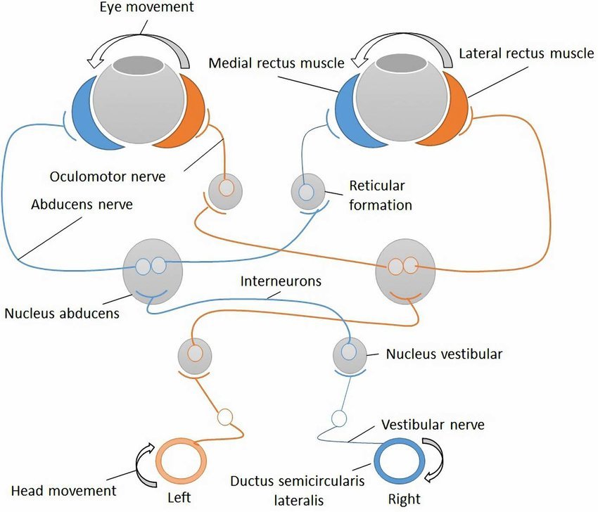
14. How does the vestibular system interact with eye movements?
A: The interaction between the vestibular system and eye movements is primarily managed by a remarkable reflex known as the Vestibulo-Ocular Reflex (VOR). Its purpose is to maintain a stable image on the retina during head movement by moving the eyes in the exact opposite direction of the head.
Without this system, your vision would appear “shaky” or blurred whenever you walked or moved your head, similar to a handheld camera being carried by someone running.
1. The Mechanics of the VOR
The VOR is one of the fastest reflexes in the human body because it is a “three-neuron arc.” This short path allows for a nearly instantaneous response (latency of less than 10 milliseconds).
- Sensing the Motion: As your head rotates (e.g., to the left), the fluid in the semicircular canals deflects the cupula.
- Signal Transmission: The vestibular nerve carries this signal to the vestibular nuclei in the brainstem.
- Eye Muscle Activation: The brainstem immediately sends signals to the extraocular muscles.
- To compensate for a leftward head turn, the reflex activates the right lateral rectus and the left medial rectus muscles.
- This pulls both eyes to the right at the exact same speed as the head movement.
2. Linear Eye Movements (Otolith-Ocular Reflex)
While the semicircular canals handle rotation, the utricle and saccule handle linear movements (like riding in a car or an elevator).
- Translational VOR: When you move sideways, your eyes move in the opposite direction to maintain focus on a target.
- Ocular Tilt Reflex: When you tilt your head toward your shoulder, the otolith organs trigger a compensatory roll of the eyes (torsional movement) to keep the vertical axis of your vision aligned with gravity.
3. Nystagmus: The “Reset” Mechanism
When head rotation is sustained or very fast, the eyes cannot move infinitely in the opposite direction; they eventually reach the limit of how far they can turn in the socket. To handle this, the brain uses nystagmus:
- Slow Phase: The eyes move smoothly in the opposite direction of the head (the VOR at work).
- Fast Phase: The eyes “flick” rapidly back to the center or in the direction of the turn to pick up a new target.
4. Visual-Vestibular Conflict
The brain constantly compares vestibular data with visual data. If these two signals don’t match, it can lead to significant distress:
Oscillopsia: A condition where the VOR is damaged. Patients feel like the world is “jumping” or “bouncing” whenever they move, because their eyes are no longer compensating for their head movements.
Motion Sickness: Occurs when your vestibular system senses motion (e.g., the rocking of a boat) but your eyes see a stable environment (the interior of a cabin).
15. What is Endolymph role in the vestibular system?
- Sensory Transduction (Mechanical-to-Electrical Conversion): This is the primary and most vital role of endolymph.
- In the Semicircular Canals: When your head rotates (angular acceleration), the inertia of the endolymph causes it to lag behind the movement of the semicircular ducts. This relative movement of the endolymph exerts pressure on a gelatinous structure called the cupula, which sits atop the hair cells. The bending of the cupula, in turn, bends the cilia (tiny hair-like projections) of the hair cells.
- In the Otolith Organs (Utricle and Saccule): When your head undergoes linear acceleration (forward/backward, up/down) or changes position relative to gravity, the otoconia (tiny calcium carbonate crystals) embedded in a gelatinous membrane within these organs shift. This shift drags the underlying gelatinous membrane, causing the cilia of the hair cells to bend.
- Electrochemical Gradient for Hair Cell Activation: The high potassium concentration in the endolymph creates a strong electrochemical gradient (an electrical potential difference) between the endolymph and the inside of the hair cells. When the hair cells’ cilia bend, specialized ion channels on their tips open, allowing a rapid influx of positively charged potassium ions from the endolymph into the hair cells. This influx causes the hair cells to depolarize (become electrically excited), leading to the release of neurotransmitters that signal the vestibular nerve and ultimately your brain about head movement and position.
16. What is the role of Perilymph in the vestibular system?
1. Physical Protection and Cushioning
The most immediate role of perilymph is structural. The membranous labyrinth is an incredibly thin, fragile “balloon” of tissue.
- Shock Absorption: Perilymph acts as a hydraulic cushion, protecting the inner ear’s sensory structures from mechanical shocks and sudden pressure changes.
- Support: It keeps the membranous structures suspended and properly positioned within the hard bone of the skull.
2. Chemical Insulation
Perilymph is chemically distinct from endolymph. It is high in Sodium ($Na^+$) and low in Potassium ($K^+$), making it very similar to extracellular fluid or cerebrospinal fluid.
- Creating the Gradient: By surrounding the membranous labyrinth, perilymph helps maintain the tight ion boundaries necessary for balance.
- The Voltage Gate: The difference in potassium concentration between the endolymph (high $K^+$) and the perilymph/hair cell body (low $K^+$) creates the electrochemical gradient that allows the sensory hair cells to fire. Without this “low-potassium” environment on the outside of the membrane, the high-potassium endolymph on the inside wouldn’t be able to drive the signal.
3. Pressure Regulation and Drainage
Perilymph is in constant communication with the Cerebrospinal Fluid (CSF) through a small channel called the cochlear aqueduct.
- Pressure Equalization: This connection helps regulate the fluid pressure within the inner ear, ensuring that changes in intracranial pressure (from coughing, straining, or altitude changes) don’t immediately damage the delicate vestibular membranes.
- Fluid Exchange: It provides a pathway for waste products to be filtered out of the inner ear space.
4. Transmission of Sound and Energy
While more critical for the cochlea (hearing), the perilymph in the vestibule also helps dissipate mechanical energy. When the stapes bone moves at the oval window, it creates pressure waves in the perilymph. This fluid displacement is what eventually allows the system to remain flexible and responsive to movement.
17. What are the main differences between Endolymph and Perilymph in the Vestibular system?
Endolymph:
- Location: Endolymph fills the membranous labyrinth itself. This includes the semicircular ducts (within the semicircular canals) and the utricle and saccule (the otolith organs).
- Composition: This is its most unique and crucial feature. Unlike most extracellular fluids in the body, endolymph is similar to intracellular fluid, meaning it is extremely high in potassium (K+) and very low in sodium (Na+). This unusual ionic composition is actively maintained by specialized cells (like the dark cells in the vestibular system and stria vascularis in the cochlea).
Perilymph:
- Location: Perilymph fills the space between the bony labyrinth (the hard, outer casing of the inner ear carved into the temporal bone) and the membranous labyrinth (the soft, fluid-filled sacs and ducts within the bony labyrinth). Think of it as a protective, cushioning fluid that surrounds the delicate membranous structures.
- Composition: Its ionic composition is similar to that of cerebrospinal fluid (CSF) and extracellular fluid, meaning it is high in sodium (Na+) and low in potassium (K+).
| Feature | Endolymph | Perilymph |
| Main Ion | Potassium (K+) | Sodium (Na+) |
| Electrical Potential | High positive potential (+80 mV) | Near zero (0 mV) |
17. What is the mechanism of VOR?
The Vestibulo-Ocular Reflex (VOR) is one of the fastest and most critical reflexes in the human body. Its primary mechanism is to maintain visual stability by rotating the eyes in the exact opposite direction of a head movement, ensuring the image remains centered on the fovea of the retina.
The VOR operates through a “three-neuron arc” that connects the inner ear directly to the eye muscles.
1. The Three-Neuron Arc
The speed of the VOR (less than 10 milliseconds) is possible because the signal only passes through three primary sets of neurons:
Step 1: The Peripheral Sensory Neuron
When the head rotates (for example, to the left), the fluid (endolymph) in the horizontal semicircular canal lags behind. This pushes the cupula, bending the hair cells in the crista ampullaris.
- The Signal: This mechanical bend is converted into an electrical signal that travels along the Vestibular Nerve (Cranial Nerve VIII).
- The Cell Body: These neurons have their cell bodies in Scarpa’s Ganglion.
Step 2: The Interneuron (Brainstem)
The vestibular nerve carries the signal into the brainstem, where it synapses in the Vestibular Nuclei (located in the pons and medulla).
- The Action: The vestibular nuclei act as the processing center, immediately “routing” the command to the specific motor nuclei responsible for moving the eyes horizontally.
Step 3: The Motor Neuron
The signal travels from the vestibular nuclei to the Ocular Motor Nuclei (Cranial Nerves III and VI).
- The Result: These neurons stimulate the extraocular muscles to pull the eyes in the opposite direction of the head turn.
2. Push-Pull Dynamics
The mechanism relies on a “push-pull” relationship between the two ears. Using a left head turn as an example:
- Left Ear (Excitation): The hair cells in the left horizontal canal are stimulated, increasing their firing rate.
- Right Ear (Inhibition): Simultaneously, the hair cells in the right horizontal canal are inhibited, decreasing their firing rate.
- Coordination: The brain compares these two signals. The difference in firing rates tells the brain exactly how fast the head is turning, allowing for a perfectly matched eye movement.
3. Muscle Synergy
To move the eyes smoothly, the reflex must coordinate multiple muscles at once. In a leftward head turn:
- The Right Lateral Rectus (pulls the right eye toward the ear) is contracted.
- The Left Medial Rectus (pulls the left eye toward the nose) is contracted.
- The opposing muscles are simultaneously inhibited to prevent resistance.
4. Gain and Phase
For the VOR mechanism to work perfectly, it must maintain two properties:
- Gain: The ratio of eye velocity to head velocity. Ideally, the gain is 1.0 (if the head moves 10° to the left, the eyes move exactly 10° to the right).
- Phase: The timing of the movement. The eyes must move at the exact same time as the head (zero latency).
Why the Mechanism Fails
If the vestibular system is damaged (e.g., vestibular neuritis), the “push-pull” balance is lost. The brain may perceive the head is turning even when it is still, leading to nystagmus (rhythmic jerking of the eyes) and a loss of visual stability during movement, known as oscillopsia.
18. How is the vestibular system connected to balance?
- Detection of Movement: The vestibular system comprises two main parts:
- Semicircular canals: These three fluid-filled loops detect rotational movements of your head (like nodding, shaking your head, or tilting it to the side). As your head moves, the fluid inside the canals shifts, bending tiny hair cells that send signals to your brain.
- Otolith organs (utricle and saccule): These organs detect linear movements (like moving forward/backward or up/down in an elevator) and the pull of gravity, informing your brain about your head’s position relative to the ground. They contain small crystals (otoconia) embedded in a gel-like membrane, which shift with movement and stimulate hair cells.
- Information Integration: The signals from the vestibular system are sent to your brain, specifically to areas like the brainstem and cerebellum. Your brain then integrates this information with input from other sensory systems, including:
- Vision: What you see helps orient you in your surroundings.
- Proprioception: Sensory information from your muscles and joints tells your brain about your body’s position and movement.
- Motor Output for Balance Control: Based on this integrated sensory information, your brain sends signals to your muscles and eyes to make rapid, automatic adjustments to maintain balance. Two key reflexes are involved:
- Vestibulo-ocular reflex (VOR): This reflex stabilizes your gaze by causing your eyes to move in the opposite direction of your head movements. This ensures your vision remains clear even when you’re moving.
- Vestibulospinal reflex (VSR): This reflex automatically adjusts your body’s posture and muscle tone to prevent you from falling, especially when you encounter uneven surfaces or changes in your position.
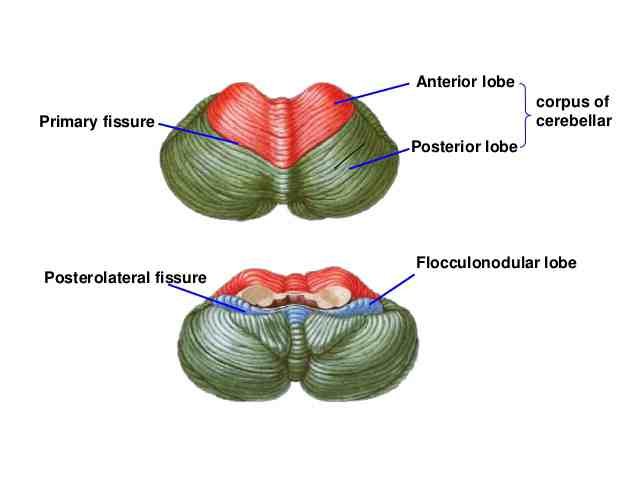
19. What is the role of the cerebellum in vestibular function?
- Integration of Sensory Information: The cerebellum is a master integrator. It receives extensive sensory input from:
- The vestibular system: Direct signals from the inner ear (semicircular canals and otolith organs) about head movement and position.
- Proprioceptors: Information from muscles, tendons, and joints throughout the body about body position and movement.
- Vision: Visual cues about the environment and self-motion.
- Motor cortex: Commands from the cerebral cortex regarding intended movements. This integration allows the cerebellum to create a comprehensive picture of the body’s current state and intended actions, crucial for effective balance control.
- Refinement of Vestibular Reflexes:
- Vestibulo-ocular Reflex (VOR) Adaptation: The VOR ensures stable vision during head movements. The cerebellum is absolutely essential for adapting and fine-tuning the VOR. If you wear new glasses that magnify or minify the world, your VOR needs to adjust so your eyes still move appropriately to keep images clear. This learning process, known as VOR adaptation, is heavily dependent on the cerebellum. It uses “error signals” (e.g., visual slip on the retina when the VOR isn’t perfectly compensating) to modify the reflex’s gain (the ratio of eye velocity to head velocity).
- Vestibulospinal Reflex (VSR) Modulation: The VSR helps maintain posture and balance by adjusting muscle tone in the trunk and limbs. The cerebellum modulates these reflexes, ensuring that postural adjustments are smooth, appropriate, and anticipatory to maintain stability. For example, if you’re about to step on an uneven surface, the cerebellum helps refine the necessary muscle adjustments to prevent a fall.
- Motor Learning and Adaptation: The cerebellum is a key player in motor learning, including the learning of new balance strategies. When you encounter a new environment (e.g., walking on ice) or learn a new motor skill (e.g., riding a bike), the cerebellum helps to adjust and optimize motor commands based on sensory feedback and predictions. It uses a trial-and-error process to refine movements and minimize errors, leading to more efficient and coordinated balance control over time.
- Coordination and Smoothness of Movement: Damage to the cerebellum leads to a characteristic set of symptoms, collectively known as ataxia.
- Predictive Control: The cerebellum is thought to generate “internal models” of the body and its interaction with the environment. These models allow the brain to predict the sensory consequences of motor commands, enabling anticipatory adjustments to posture and eye movements even before sensory feedback arrives. This predictive capability is crucial for maintaining dynamic balance.
20. What is the macula in the vestibular system?
A: The macula is the specialized sensory tissue located within the two otolith organs of the inner ear: the utricle and the saccule. While the semicircular canals use the crista ampullaris to detect rotation, the macula is designed to detect linear acceleration and the pull of gravity.
Structure of the Macula
The macula is a complex, multi-layered patch of tissue that acts as a “gravity sensor.” It consists of three primary layers:
- Sensory Hair Cells: These are the foundation of the macula. They feature bundles of small hairs (stereocilia) and one large hair (kinocilium).
- Otolithic Membrane: A thick, gelatinous layer that sits directly on top of the hair cells. The “hairs” of the sensory cells are embedded into this gel.
- Otoconia: Thousands of microscopic calcium carbonate crystals (the “ear stones”) that sit on the surface of the gelatinous membrane.
How it Functions
The macula operates through the principle of inertia and mass:
- Adding Weight: The otoconia make the otolithic membrane significantly heavier than the surrounding fluid (endolymph).
- Linear Acceleration: When you move in a straight line (like a car accelerating or an elevator rising), the heavy membrane lags behind the movement of the head. This “lag” pulls the gelatinous layer, which bends the embedded hair cells.
- Static Tilt (Gravity): When you tilt your head, gravity pulls the weighted membrane “downhill.” This provides a constant signal to the brain about the head’s position relative to the ground, even if you aren’t moving.
Differences in Orientation
There are two maculae in each ear, oriented in different planes to cover 3D space:
- Macula Utriculi (Utricle): Located on the “floor” of the utricle. It is oriented horizontally and detects side-to-side or forward-backward movement, as well as lateral head tilts.
- Macula Sacculi (Saccule): Located on the “wall” of the saccule. It is oriented vertically and detects up-and-down movement (like an elevator) and forward/backward head tilts.
The Striola
Each macula is divided by a curved central line called the striola. The hair cells are organized in a mirrored fashion across this line. This arrangement ensures that regardless of which way the head tilts or moves, some hair cells will be excited while others are inhibited, allowing the brain to pinpoint the exact direction of motion.
21. Why are there two vestibular systems (one in each ear)?
A: Having bilateral vestibular systems allows the brain to compare input from both ears, enabling accurate detection of direction and magnitude of head movement, enhancing balance and orientation.
22. What happens when the vestibular system is damaged?
A: The effects can be categorized into sensory, physical, and cognitive symptoms.
1. Sensory Symptoms: Vertigo and Dizziness
The hallmark of vestibular damage is a mismatch in the “push-pull” dynamic between the two ears.
- Vertigo: A false sensation of movement. If one vestibular nerve is damaged (e.g., Vestibular Neuritis), the brain perceives the constant firing from the healthy ear as a continuous turn, causing the room to feel like it is spinning.
- Nystagmus: Because of the Vestibulo-Ocular Reflex (VOR), the brain tries to compensate for the perceived “turn” by moving the eyes. This results in a rhythmic, involuntary jerking of the eyes as they drift slowly one way and “flick” back to center.
2. Visual Symptoms: Loss of Gaze Stability
When the VOR is impaired, the eyes no longer move in perfect opposition to the head.
- Oscillopsia: This is the sensation that the world is “bouncing” or “jumping” whenever the person moves. It occurs because the eyes are not staying fixed on a target while the head is in motion (walking, driving, or even just turning the head).
- Blurred Vision: Vision may become blurry during movement because the brain cannot stabilize the image on the retina quickly enough.
3. Physical Symptoms: Imbalance and Falls
Damage to the otolith organs (utricle and saccule) or their pathways affects the vestibulospinal reflex, which controls posture.
- Ataxia: A lack of muscle coordination while walking. A person with vestibular damage may walk with a “wide-based gait” or lean heavily toward the side of the damaged ear.
- Postural Instability: Difficulty standing upright, especially in the dark or on uneven surfaces (like grass or sand), where the brain cannot rely as heavily on vision or touch.
4. The “Brain Fog” and Autonomic Response
The vestibular system is closely linked to the area postrema (the brain’s vomiting center) and higher cognitive centers.
- Nausea and Vomiting: The sensory conflict between the eyes and the ears often triggers the same response as motion sickness.
- Cognitive Fatigue: Because the brain has to work much harder to consciously maintain balance—a task that is normally automatic—patients often experience extreme fatigue, “brain fog,” and difficulty concentrating.
5. Specific Conditions
The symptoms often depend on the nature of the damage:
Bilateral Vestibular Loss: When both sides are damaged (often by certain ototoxic medications), the person may not experience “spinning” but will have severe trouble walking and debilitating oscillopsia.
BPPV (Benign Paroxysmal Positional Vertigo): Caused by otoconia (crystals) falling into the semicircular canals, leading to brief but intense spinning when changing head position.
Meniere’s Disease: Caused by an over-accumulation of endolymph, resulting in fluctuating hearing loss, tinnitus, and episodes of vertigo.
23. How do semicircular canals achieve directional sensitivity?
A: This system allows the brain to determine exactly which direction the head is turning by comparing the “data stream” from all six canals simultaneously.
1. Orthogonal Orientation (The XYZ Axes)
The primary way the canals achieve directional sensitivity is through their physical layout. In each ear, the three canals are oriented roughly 90 degrees to one another, mimicking a 3D coordinate system ($x$, $y$, and $z$ axes):
- Horizontal (Lateral) Canal: Tilted back about 30 degrees from the horizontal plane; it detects rotation around a vertical axis (e.g., shaking your head “no”).
- Anterior (Superior) Canal: Detects rotation in the sagittal plane (e.g., nodding “yes”).
- Posterior Canal: Detects rotation in the frontal plane (e.g., tilting your head toward your shoulder).
2. Functional Pairing (Push-Pull Dynamics)
The canals in the left ear are mirrored by the canals in the right ear. They work in “functional pairs” located in the same geometric plane. When one canal in a pair is stimulated, its partner on the other side of the head is inhibited.
- The Pairs:
- Left Horizontal and Right Horizontal.
- Left Anterior and Right Posterior.
- Left Posterior and Right Anterior.
How this signals direction:
If you turn your head to the left, the fluid in the left horizontal canal moves toward the sensory organ (excitation), while the fluid in the right horizontal canal moves away from it (inhibition). The brain compares these opposing signals to confirm the direction and speed of the turn.
3. Morphological Polarization
Directional sensitivity at the cellular level is determined by the arrangement of the hair cells within the crista ampullaris.
Each hair cell has a single, tallest hair called the kinocilium. The direction in which the hair bundle is bent determines the signal sent to the brain:
- Excitation: Bending the hairs toward the kinocilium opens ion channels, causing the nerve to fire more rapidly.
- Inhibition: Bending the hairs away from the kinocilium closes channels, slowing the firing rate.
4. Directional Specificity by Canal
The hair cells are arranged differently depending on which canal they are in:
Vertical Canals (Anterior/Posterior): The kinocilia are positioned away from the utricle (ampullofugal flow). Therefore, fluid moving away from the ampulla causes excitation.
Horizontal Canals: The kinocilia are positioned toward the utricle (ampullopetal flow). Therefore, fluid moving toward the ampulla causes excitation.
QUIZ
1. Which of the following structures is part of the vestibular system?
A. Cochlea
B. Semicircular canals
C. Ossicles
✅ Correct Answer: B. Semicircular canals
2. How many semicircular canals are present in total (both ears)?
A. 2
B. 3
C. 6
✅ Correct Answer: C. 6
3. What type of head movement do the semicircular canals detect?
A. Linear acceleration
B. Sound waves
C. Angular (rotational) acceleration
✅ Correct Answer: C. Angular (rotational) acceleration
4. What is the function of the otolith organs?
A. Detect sound
B. Detect rotational head movements
C. Detect linear acceleration and gravity
✅ Correct Answer: C. Detect linear acceleration and gravity
5. The utricle primarily detects:
A. Rotational movement
B. Vertical movement
C. Horizontal linear acceleration
✅ Correct Answer: C. Horizontal linear acceleration
6. What are otoconia composed of?
A. Fatty acids
B. Potassium ions
C. Calcium carbonate crystals
✅ Correct Answer: C. Calcium carbonate crystals
7. What fluid fills the membranous labyrinth?
A. Perilymph
B. Endolymph
C. Plasma
✅ Correct Answer: B. Endolymph
8. Hair cells transduce mechanical signals into:
A. Light signals
B. Thermal signals
C. Electrical signals
✅ Correct Answer: C. Electrical signals
9. What reflex keeps your vision stable during head movement?
A. Startle reflex
B. Acoustic reflex
C. Vestibulo-ocular reflex (VOR)
✅ Correct Answer: C. Vestibulo-ocular reflex (VOR)
10. The macula is found in:
A. Cochlea
B. Ampulla
C. Utricle and saccule
✅ Correct Answer: C. Utricle and saccule
PHYSIOLOGY OF VESTIBULAR SYSTEM
1. What is the primary function of the semicircular canals?
1. Detection of Rotation
The three canals (anterior, posterior, and horizontal) are positioned at approximately right angles to one another. This “XYZ” alignment allows the system to detect rotation in any direction:
- Horizontal Canal: Detects side-to-side rotation (e.g., shaking your head “no”).
- Anterior Canal: Detects forward and backward motion (e.g., nodding “yes”).
- Posterior Canal: Detects lateral tilting (e.g., tipping your ear toward your shoulder).
2. Maintaining Visual Stability (The VOR)
One of the most critical roles of the semicircular canals is to drive the Vestibulo-Ocular Reflex (VOR).
- As the canals sense the head rotating in one direction, they send immediate signals to the eye muscles to move the eyes in the opposite direction.
- Function: This keeps your gaze fixed on a target even while your head is moving, preventing your vision from becoming blurry or “shaky.”
3. Coordinating Balance and Posture
The canals send constant data to the brainstem and cerebellum. The brain uses this information to:
- Identify the speed and direction of a turn.
- Anticipate changes in body position.
- Trigger “righting reflexes” in the trunk and limbs to prevent you from losing your balance during complex movements, like turning a corner while walking.
How They Work (In Brief)
The canals achieve this through fluid dynamics:
The bending of the cupula triggers sensory hair cells, which send electrical impulses through the vestibular nerve to the brain.
2. What is the semicircular canals mechanism of action?
When your head rotates, the bony labyrinth moves, but the endolymph inside the membranous semicircular canals lags behind due to inertia.
This relative movement of the endolymph pushes against the cupula.
The bending of the cupula deflects the stereocilia (and kinocilium, in vestibular hair cells) of the hair cells within the crista ampullaris.
These bending opens or closes mechanically gated ion channels in the hair cells, leading to depolarization or hyperpolarization.
The change in electrical potential alters the release of neurotransmitters at the synapse with the vestibular nerve fibers (part of the VIII cranial nerve).
These nerve fibers transmit signals to the brainstem, where the information is processed to give you the sensation of rotation and trigger compensatory reflexes like the vestibulo-ocular reflex (VOR), which stabilizes your gaze during head movements.
3. What type of fluid is found inside the semicircular canals?
A: The fluid found directly inside the semicircular canals (within the membranous labyrinth) is called endolymph.
Characteristics of Endolymph
Endolymph is unique because its chemical composition is unlike most other extracellular fluids in the body. It is often compared to intracellular fluid (the fluid found inside cells) due to its high concentration of ions.
- High Potassium ($K^+$): This is the most critical feature. The high potassium levels create an electrochemical gradient that allows for the rapid firing of sensory hair cells.
- Low Sodium ($Na^+$): In most other body fluids, sodium is the dominant ion, but in endolymph, it is kept at very low levels.
- Positive Potential: Endolymph has a significant positive electrical potential (about $+80$ mV), which acts like a “battery” to power the hair cells.
How It Works in the Canals
The endolymph functions as a mechanical messenger. When your head rotates:
- The bony canals move with your head.
- The endolymph inside lags behind due to inertia.
- This lagging fluid pushes against the cupula (the gelatinous “sail” in the ampulla).
- The bending of the cupula triggers the hair cells to send a signal to the brain.
The “Other” Fluid: Perilymph
It is important to note that while endolymph is inside the canals, another fluid called perilymph surrounds the outside of the membranous tubes, acting as a cushion between the membrane and the bone of the skull.
| Feature | Endolymph | Perilymph |
| Location | Inside the membranous labyrinth | Between bone and membrane |
| Dominant Ion | Potassium ($K^+$) | Sodium ($Na^+$) |
| Function | Sensory signal transduction | Protection and cushioning |
4. What are the main differences between Endolymph and Perilymph?
Feature
Endolymph
Perilymph
Location
Within the membranous labyrinth (cochlear duct, semicircular canals, utricle, saccule)
Between the bony labyrinth and the membranous labyrinth (scala vestibuli, scala tympani)
Ionic Composition
High in potassium (K+), low in sodium (Na+) and calcium (Ca2+)
High in sodium (Na+), low in potassium (K+) and calcium (Ca2+)
Origin
Secreted by the stria vascularis and dark cells
Derived from blood plasma and cerebrospinal fluid (CSF)
Electrical Potential
Positive (+80 to +90 mV relative to perilymph)
Negative (0 mV relative to itself)
Function
Plays a vital role in the excitation of hair cells for sound and vestibular transduction
Transmits vibrations from the oval window and provides a fluid environment for the membranous labyrinth
Similarity to
Intracellular fluid
Extracellular fluid, CSF
When your head rotates, the bone of the canal moves, but the fluid inside (endolymph) lags behind due to inertia.
This lagging fluid pushes against a gelatinous “sail” called the cupula.
5. Which part of the semicircular canal bends in response to fluid movement, activating hair cells?
A: When the head rotates in the plane of a specific semicircular canal:
The bony labyrinth and the semicircular duct move with the head.
The endolymph, due to its inertia, tends to lag behind.
This relative movement of the endolymph causes it to exert pressure against the cupula.
The cupula then bends (or deflects) in the direction of the endolymph flow.
The bending of the cupula, in turn, causes the embedded stereocilia of the hair cells to bend.
This mechanical bending of the stereocilia opens ion channels in the hair cell membrane, leading to a change in the hair cell’s electrical potential (depolarization or hyperpolarization).
This change in electrical potential triggers the release of neurotransmitters, which then generate signals in the afferent nerve fibers of the vestibular nerve, sending information about the head’s rotation to the brain.
6. The orientation of the semicircular canals allows the detection of motion in which directions?
A: The semicircular canals are oriented in three distinct planes, roughly 90° to each other, functioning like a 3D coordinate system (x,y, and z axes). This anatomical layout allows the vestibular system to detect angular acceleration (rotation) in any direction.
1. The Horizontal (Lateral) Canal
This canal is responsible for detecting rotation in the transverse plane.
- Motion detected: Side-to-side rotation around a vertical axis.
- Example: Shaking your head “no” or spinning in a swivel chair.
- Note: Anatomically, it is tilted back about 30° relative to the true horizontal plane of the earth.
2. The Anterior (Superior) Canal
This canal detects motion in the sagittal plane.
- Motion detected: Forward and backward rotation around a frontal-horizontal axis.
- Example: Nodding your head “yes” or performing a somersault.
3. The Posterior Canal
This canal detects motion in the frontal (coronal) plane.
- Motion detected: Lateral rotation around a sagittal-horizontal axis.
- Example: Tilting your head toward your shoulder or performing a cartwheel.
Functional Pairing
The canals work in mirrored “push-pull” pairs. Because of their specific angles, a movement that excites a canal in one ear will inhibit its functional partner in the other:
Right Anterior + Left Posterior: Paired for diagonal/nodding movements.
Left Horizontal + Right Horizontal: Paired for “no” movements.
Left Anterior + Right Posterior: Paired for diagonal/nodding movements.
By integrating the signals from all three planes simultaneously, the brain can calculate complex, multi-axis rotations (such as looking up and to the right while walking).
7. What type of motion every single semicircular canal detects?
Horizontal (or Lateral) Semicircular Canal:
Orientation: Lies in an approximately horizontal plane when the head is tilted forward by about 30 degrees (which is the typical position of the head when standing upright and looking straight ahead).
Motion Detected: Primarily detects rotational movements around a vertical axis, like shaking your head “no” (yaw axis rotation).
Anterior (or Superior) Semicircular Canal:
Orientation: Lies in a vertical plane, oriented diagonally (about 45 degrees from the sagittal and frontal planes).
Motion Detected: Primarily detects rotational movements in the sagittal plane, like nodding your head “yes” (pitch axis rotation), or somersaulting forward.
Posterior (or Inferior) Semicircular Canal:
Orientation: Also lies in a vertical plane, oriented diagonally and roughly perpendicular to the anterior canal.
Motion Detected: Primarily detects rotational movements in the frontal plane, like tilting your head towards your shoulder (roll axis rotation), or doing a cartwheel.
8. How does Semicircular Canal pairing system work?
A: The Semicircular Canal pairing system, often referred to as the Push-Pull dynamic, is the clever anatomical arrangement that allows your brain to precisely track head rotation in 3D space. It relies on the fact that your six canals (three in each ear) are organized into three mirrored, functional pairs.
1. The Three Functional Pairs
Each canal in your left ear sits in the same geometric plane as a specific canal in your right ear. When you move your head in that plane, both canals are activated—but in opposite ways.
| Functional Pair | Planes of Motion |
| Left & Right Horizontal | Horizontal rotation (e.g., shaking your head “no”). |
| Left Anterior & Right Posterior (LARP) | Diagonal nodding (e.g., looking down at your left shoe). |
| Right Anterior & Left Posterior (RALP) | Diagonal nodding (e.g., looking down at your right shoe). |
2. The “Push-Pull” Mechanism
The pairing system works because the sensory hair cells in mirrored canals are oriented in opposite directions.
- Excitation (The Push): When you turn your head, the fluid (endolymph) in the canal toward the direction of the turn pushes against the sensory organ (cupula), causing the nerve to fire faster.
- Inhibition (The Pull): Simultaneously, the fluid in the mirrored canal in the other ear moves away from its sensory organ, causing that nerve to fire slower.
Example: Turning your head to the Left
- Left Horizontal Canal: Detects an “ampullopetal” flow (fluid moving toward the ampulla), which increases the firing rate.
- Right Horizontal Canal: Detects an “ampullofugal” flow (fluid moving away), which decreases the firing rate.
- The Brain: Sees one side go “Up” and the other go “Down.” This difference allows the brain to confirm the direction and speed of the turn with high resolution.
and Why is this pairing necessary?
The pairing system provides three major biological advantages:
- Accuracy: By comparing two data points (one increasing, one decreasing) rather than just one, the brain can filter out “background noise” and detect even very tiny head movements.
- Redundancy: If one ear is damaged or weakened, the brain can still rely on the “pull” (inhibition) from the healthy side to figure out which way the head is moving.
- Sensitivity to High Speed: According to Ewald’s Second Law, our ears are better at sensing “excitation” than “inhibition.” Having a pair ensures that no matter which way you turn, one ear is always providing that high-intensity excitatory signal.
Clinical Relevance
When this pairing system is disrupted, it leads to significant balance issues. If one nerve is damaged (e.g., by a virus), the brain receives a “low” signal from one side and a “normal” signal from the other. Even if you are standing perfectly still, the brain interprets this asymmetry as a constant head rotation, resulting in the spinning sensation of vertigo.
9. What does the Push Pull system mean?
A: The “push-pull” system means that when the head rotates in a specific plane, one canal in the pair is excited (increased firing rate) while its partner is inhibited (decreased firing rate). This complementary signaling provides the brain with precise information about the direction and speed of head rotation.
10. What is Ampulla?
A: An ampulla (plural: ampullae) is a bulbous or dilated enlargement at the base of each of the three semicircular canals.
Location of Sensory Organs: The ampulla is crucial because it houses the primary sensory organ for detecting angular acceleration (rotational head movements). This sensory structure is called the crista ampullaris.
Crista Ampullaris: This is a ridge or mound of specialized tissue within the ampulla. It contains the actual hair cells, which are the mechanoreceptors responsible for converting mechanical movement into electrical signals.
Cupula: Overlying the crista ampullaris is a gelatinous, dome-shaped structure called the cupula. The “hairs” (stereocilia and kinocilium) of the hair cells are embedded within this cupula, and the cupula extends across the entire lumen (opening) of the ampulla, forming a barrier to the endolymph.
111. What does Ampulla do?
A: It plays a crucial role in detecting rotational (angular) movements of the head.
This is how:
When the head rotates, the endolymph (the fluid inside the semicircular canal) lags behind due to inertia.
This relative movement of the endolymph exerts pressure on the cupula.
The cupula then bends (deflects) in the direction of the endolymph flow.
This bending of the cupula, in turn, bends the embedded stereocilia of the hair cells.
The bending of the hair cells’ stereocilia causes changes in their electrical potential, leading to the generation of nerve impulses that are sent to the brain via the vestibular nerve.
Left Horizontal + Right Horizontal: Paired for “no” movements.
Left Anterior + Right Posterior: Paired for diagonal/nodding movements.
12. How do Utricle and Saccule react to head tilt-Static Equilibrium?
When you tilt your head, gravity acts on the heavy otoconia.
The weight of the otoconia causes the otolithic membrane to shift or slide relative to the macula.
This shearing motion bends the stereocilia of the hair cells embedded within the gelatinous layer.
Depending on the direction of the bend (towards or away from the kinocilium), the hair cells either depolarize (increase firing rate) or hyperpolarize (decrease firing rate).
This change in electrical activity generates nerve impulses that are sent to the brain via the vestibular nerve (specifically, the superior vestibular nerve for the utricle and the inferior vestibular nerve for the saccule).
The brain interprets these signals to understand your head’s static position relative to gravity (e.g., whether your head is upright, tilted forward, or tilted to the side).
13. How do Utricle and Saccule react to linear acceleration-Dynamic Equilibrium?
When your head undergoes linear acceleration (e.g., starting to walk forward, accelerating in a car, or going up/down in an elevator), the otoconia, due to their inertia, lag behind the movement of the head and the macula.
This inertial lag causes the otolithic membrane to slide or shear across the hair cells.
Again, this shearing motion bends the hair cells’ stereocilia, leading to changes in their electrical activity and the generation of nerve impulses.
The brain interprets these signals as linear acceleration (e.g., knowing you’re moving forward, backward, up, or down).
14. What is the Stereocilia function in the vestibular system?
I. SemicircularCanals (Rotational Motion):
Movement of the endolymph in the semicircular canals deflects the cupula (the gelatinous cap in the ampulla).
The bending of the cupula, in turn, bends the stereocilia of the hair cells embedded within it.
Bending the stereocilia towards the kinocilium (in vestibular hair cells) depolarizes the cell, increasing nerve firing.
Bending them away from the kinocilium hyperpolarizes the cell, decreasing nerve firing. This directional sensitivity is key to detecting rotational direction.
Otolith Organs (Utricle and Saccule – Linear Motion & Gravity):
The weight and inertia of the otoconia (calcium carbonate crystals) embedded in the gelatinous layer overlying the hair cells cause the otolithic membrane to shift during linear acceleration or head tilt.
This shearing motion directly bends the stereocilia.
Similar to the canals, the direction of bending relative to the kinocilium determines whether the hair cell depolarizes or hyperpolarizes, signaling the brain about linear movement or static head position relative to gravity.
15. What is the kinocilium function in the vestibular system?
I. Establishes Hair Cell Polarity:
Each vestibular hair cell has a single kinocilium, which is typically the tallest cilium in the hair bundle.
The kinocilium is positioned at one end of the staircase-like arrangement of stereocilia. This specific anatomical orientation defines the morphological polarity of the hair cell.
This polarity dictates the direction in which the hair bundle needs to be bent to either excite (depolarize) or inhibit (hyperpolarize) the hair cell.
II. Directional Sensitivity and Signal Modulation:
Bending of stereocilia towards the kinocilium: This causes tension on the tip links (which connect the stereocilia), mechanically opening ion channels. The influx of positive ions (primarily potassium) into the hair cell leads to depolarization (excitation) and an increase in the firing rate of the associated vestibular nerve fiber.
Bending of stereocilia away from the kinocilium: This releases the tension on the tip links, causing the ion channels to close. This prevents ion influx, leading to hyperpolarization (inhibition) and a decrease in the firing rate of the vestibular nerve fiber.
Mechanical Link and Force Transmission:
It acts as a mechanical lever or pivot. In the semicircular canals, it is embedded in the cupula. In the otolith organs, it is embedded in the otolithic membrane. This structural connection ensures that the forces generated by fluid movement or gravity are effectively transmitted to the stereocilia.
QUIZ
1. What is the main function of the semicircular canals?
A. Detect linear acceleration
B. Detect angular acceleration
C. Control balance through hearing
✅ Correct Answer: B
2. Which fluid is found inside the membranous semicircular canals?
A. Perilymph
B. Cerebrospinal fluid
C. Endolymph
✅ Correct Answer: C
3. Which part of the semicircular canal contains the sensory organ that detects rotation?
A. Ampulla
B. Saccule
C. Macula
✅ Correct Answer: A
4. What is the name of the structure inside the ampulla that contains the hair cells?
A. Otoconia
B. Crista ampullaris
C. Organ of Corti
✅ Correct Answer: B
5. What is the gelatinous structure that covers the hair cells in the ampulla?
A. Cupula
B. Tectorial membrane
C. Otolithic membrane
✅ Correct Answer: A
6. The semicircular canals are most sensitive to which type of movement?
A. Gravity
B. Angular or rotational movement
C. Sound pressure waves
✅ Correct Answer: B
7. The horizontal semicircular canal detects motion in which direction?
A. Side to side (yaw)
B. Up and down
C. Forward and backward
✅ Correct Answer: A
8. The anterior semicircular canal is primarily sensitive to what type of motion?
A. Nodding “yes”
B. Tilting head to shoulder
C. Shaking “no”
✅ Correct Answer: A
9. What causes the cupula to bend during head rotation?
A. Gravity acting on otoconia
B. Endolymph lag due to inertia
C. Sound waves entering the cochlea
✅ Correct Answer: B
10. The crista ampullaris responds to which type of acceleration?
A. Angular
B. Static
C. Linear
✅ Correct Answer: A
11. The otolith organs detect:
A. Rotational head movement
B. Static and dynamic linear acceleration
C. Pitch changes in sound
✅ Correct Answer: B
12. What happens when stereocilia bend toward the kinocilium?
A. Depolarization
B. Hyperpolarization
C. No response
✅ Correct Answer: A
13. Which of the following best describes endolymph?
A. High in potassium, low in sodium
B. Same composition as plasma
C. High in sodium, low in potassium
✅ Correct Answer: A
14. What structure provides the mechanical stimulus for hair cell deflection in the otolith organs?
A. Cupula
B. Otolithic membrane with otoconia
C. Tympanic membrane
✅ Correct Answer: B
15. Where is the cupula located?
A. B. Inside the saccule
B. Within the cochlear duct
C. Over the crista ampullaris in the ampulla
✅ Correct Answer: C